
Evaluation of ankle conventional x-ray as a gold standard on diagnosis and treatment decisions for ankle weber B fractures. Self-controlled multi-centric experiment.
Publicado en: Herrera-Arbelaez JM et al. Evaluation of ankle conventional x-ray as a gold standard on diagnosis and treatment decisions for ankle weber B fractures. Self-controlled multi-centric experiment. Rev Col Or Tra. 2020;34:212-22 DOI: 10.1016/j.rccot.2020.07.002
How was this research conducted?
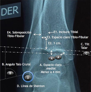
Following a review of a cohort of patients with Weber B ankle fracture-dislocations—in which we compared lateral plates versus antiglide plates over a five-year follow-up period (2012)—our research group noted that, despite finding significant differences favoring antiglide plates, there were still suboptimal outcomes in both the antiglide and lateral plate groups. Although we never identified a diagnostic error, we did observe significant discrepancies in decision-making among the various orthopedists. We therefore decided to investigate whether ankle radiography was truly the gold standard for making therapeutic decisions. We first conducted a study involving Colombian orthopedists and foot surgeons, finding a 50% discrepancy in decision-making when they evaluated only the three standard views (AP, lateral, and mortise/internal rotation); basing decisions solely on radiographs of the fractured ankle is akin to a coin toss. This discrepancy dropped to 25% when the radiographic series of the healthy contralateral ankle was also evaluated, allowing for a comparison of conventional radiological measurements between the fractured and healthy ankles. Concerned that word might spread internationally that Colombians did not know how to interpret ankle radiographs, we decided to replicate the study in Ecuador and Mexico, where we found and validated the exact same results. Of course, in none of the three countries was the fracture itself missed (100% accuracy); we therefore concluded that, for the purpose of diagnosis, the radiographic series of the fractured ankle alone is indeed the gold standard. However, radiographs cannot be considered the gold standard for making therapeutic decisions, as—even with conventional measurements and a healthy contralateral image—there is only about 75% agreement among orthopedists and/or foot and ankle surgeons. Therefore, we recommend using computed tomography (CT) with 3D visualization of the fracture to guide therapeutic decision-making.
Why is this research important?
This is the Colombian scientific paper with the highest number of downloads in the history of orthopaedic publications produced entirely in Colombia (nearly 300,000). Yet beyond the volume of readership, this publication brought about a conceptual shift, establishing that ankle fracture-dislocations should henceforth be analyzed in three dimensions using CT scans. Furthermore, it raised awareness regarding the intra-articular nature and complexity of ankle fracture-dislocations.
This scientific paper is aimed at professionals in orthopedics, emergency medicine, sports medicine, physiatry, physical therapy, and kinesiology, given that ankle fractures require the kind of "transdisciplinary" management discussed here.